There are four types of influenza viruses – A, B, C and D. Types A and B are responsible for the annual influenza epidemics that occur in humans, and type A viruses are the only influenza viruses known to cause pandemics. The general requirement for an influenza virus to cause a pandemic is for the virus to be sufficiently different from prior circulating viruses such that there is little to no immunity in the population. Influenza C viruses cause such mild illness in humans that we rarely identify those infections and we do not consider influenza C viruses to pose a pandemic threat. Influenza D viruses primarily infect cattle and can spillover to other animals, but pose little threat to humans.
Influenza viruses are RNA viruses and thus are prone to replication errors and the accumulation of mutations. Under normal circumstances, these mutations are incremental and do not result in wholly antigenically distinct viruses (meaning that our immune systems would not recognize them). This is referred to as antigenic drift, and it happens frequently enough that we experience yearly epidemics during the flu season. Antigenic drift involves minor changes in the hemagglutinin (HA) and neuraminidase (NA) proteins of the circulating influenza A viruses, such that it is necessary for us to update the influenza vaccines every year, however, not so significant of changes that some degree of cross-reactive immunity would not exist or that a new subtype of virus would emerge (the latter developments would represent antigenic shift, as opposed to drift).
Subtypes of influenza A viruses are characterized by their hemagglutinin and neuraminidase protein characteristics. There are 18 known subtypes of hemagglutinin proteins that are represented by the letter H followed by a number (H1 – H18), and 11 known subtypes of neuraminidase proteins that are represented by the letter N followed by a number (N1 – N11). The subtypes of influenza A viruses are then described by the combination of the hemagglutinin and neuraminidase subtypes. The influenza A subtypes that contribute to our seasonally recurring flu seasons are H1N1 or A(H1N1) and H3N2 or A(H3N2).
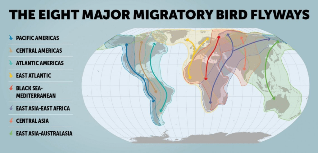
All known influenza A subtypes exist in aquatic birds, which serves as the reservoir for influenza A viruses. These aquatic birds are migratory birds and they can carry and spread the virus along their flyways.
In wild ducks, these viruses replicate in the cells lining their gastrointestinal tracts, but do not make the ducks sick. The ducks excrete very high levels of the virus and can contaminate fresh water ponds, lakes and rivers, as well as land that they fly over or dwell on, which in turn often leads to infections of domestic birds and poultry. Large numbers of baby ducks are hatched each year, and they can be infected by virus in the water, and further contribute to the spread of the virus.
It is believed that all mammalian influenza viruses derive from the aquatic bird (avian) influenza reservoir, and phylogenetic analyses (the study of the evolution of genetic sequences) suggests that both swine and human influenza A viruses evolved from avian influenza viruses.
Besides accumulating mutations as a result of errors in transcription (the process of copying the virus’ genetic material during virus replication in order to make copies of the virus’ proteins, which in turn can be assembled into new virus progeny), influenza viruses can undergo the process of reassortment.
The diagram above shows one such example that lead to the H7N9 virus that infected humans in China back in 2013.
The influenza virus has eight genes coding for eight influenza A proteins (PA, PB1, PB2, nucleoprotein, hemagglutinin, neuraminidase, nuclear export protein and membrane protein. If a host is infected with two or more different influenza viruses, these viruses can exchange genes resulting in a new virus that has genes from more than one influenza virus. In the above example, the H7N9 virus got its hemagglutinin protein H7 from a domestic duck and its neuraminidase protein N9 from wild birds, while the other genes came from multiple H9N2 viruses in domestic poultry. The significance of this is that the new resulting virus can have much more antigenic variation from that of previously circulating influenza viruses (antigenic shift) and the new collection of proteins may confer biological changes to the virus that might result in enhanced transmission to humans, and even more concerningly, the capability for enhanced forward transmission (human-to-human spread).
The first human influenza virus was isolated in 1933. The largest influenza pandemic in just over 100 years was the Spanish flu pandemic of 1918 – 1919. That was an H1N1 virus, and there is evidence to suggest that it was an intact avian influenza virus (not the result of reassortment) that appeared in humans or swine before 1918 and then replaced the previously circulating strains when it caused the pandemic.
The Asian influenza virus (H2N2) replaced the 1918 H1N1 virus in 1957 when it caused a pandemic. The Hong Kong influenza virus (H3N2) appeared in 1968 when it sparked a pandemic, and then the H1N1 virus reappeared in 1977.
In my post last night on avian influenza, I mentioned a recently published article (yesterday) that I thought should give us pause about many of the assumptions being made about the current H5N1 outbreak in dairy cattle, and that should cause us to increase and improve our response to containing this outbreak. Today, we will review that study in greater detail.
In this study, the investigators examined the H5N1 virus circulating in dairy cattle that had infected a farm worker. This is very important because we need to know whether this was just an isolated occurrence that happened by chance related to the closeness of the farm worker and length of time working with these infected animals that represents a very low risk to the general population that is not at such occupational risk, or alternatively, whether the virus is evolving in such a way as to be able to more efficiently spread to humans, and even more concerning would be if the virus is developing more effective transmission from infected humans to their human close contacts. Thus far, we have seen no evidence of the latter, which would be the most concerning as to risk to the general population and as to pandemic potential.
Somewhat surprising (at least to me) was that the virus could grow well on human lung cells (alveolar epithelial cells), but it did not grow well in eye cells (corneal epithelial cells) – both cells have the receptor type used by avian influenza viruses [my surprise being in part because the most common presentation of infected farm workers has been conjunctivitis, an infection of the lining over the eye].
We don’t test these viruses on human subjects for ethical reasons, so animal models are used, including mice. However, ferrets are one of the best animal models because they most closely mirror influenza infections in humans, so they were used in this study, as well.
When directly infected with the virus recovered from the farm worker’s eye, the infection caused rapid and deadly infections in the mice and ferrets, in fact, all of the ferrets died. This tells me that we may very well be lulled into thinking that the current H5N1 only causes mild illness in humans since that is what we have observed in the 36 humans that are known to have been infected thus far. But we need to keep in mind that is likely only because it infected their eyes and not their lungs. Our upper airways have different receptors that are not amenable to H5N1 binding, so the virus is not finding a way to get to our lungs just yet. However, to me, this means we must contain the spread of this infection in cattle and other animals so that the virus does not continue to evolve in such a way as to be able to infect our upper airways and eventually our lungs.
Another very concerning finding was that the recovered H5N1 virus was able to spread (though not terribly efficiently) through the air 17 – 33% of the time from directly infected ferrets to healthy ferrets in a separate, but nearby cage in the same room. Five of the six infected ferrets who became infected from these respiratory droplets died. This is concerning because the USDA has been telling us that cow-to-cow and cattle-to-poultry transmission is likely occurring predominantly through fomites (virus on inanimate objects such as workers clothing or the equipment used to milk the cows) and not through respiratory means, and it is further concerning, because even though not highly transmissible through respiratory droplets, this represents an increase in transmissibility compared to prior strains of the bovine H5N1 virus.
We know from our prior studies of avian influenza viruses that while avian influenza viruses generally only cause sporadic infections in mammals, including humans, that a mutation named PB2-E627K (the PB2 is a designation for one of the H5N1 proteins, the E and K specify the amino acid substitution (lysine) that is present in the protein, and the 627 specifies the exact location in the amino acid sequence of the protein where the mutation occurs. This mutation has been known to increase transmissibility of avian influenza viruses in mammals, increase the polymerase (the enzyme necessary to replicate (make more of) the virus’ proteins) activity, and to increase virulence. Thus, there is concern that the H5N1 virus circulating cattle might be evolving to better infect mammals, including humans.
Another mutation was also observed – PB2-M631L. This mutation increased polymerase activity in human cells, and this is another concerning indicator of potential evolution of the virus to adapt to enhanced mammalian transmission.
Fortunately, the mutations have not resulted in resistance to all of our available antiviral medications for influenza A, although the virus has become less sensitive to the antiviral medication that has been our standard treatment for these patients.
My take-a-way from this research is that we are likely underestimating the risks of the current outbreak of H5N1 among dairy cattle in the U.S. both in terms of the potential for spread to humans and the risk for severe disease. This does not mean that either of these are imminent, but there is a progression of warning signs that I look for with any outbreak of a novel virus.
The threshold question is whether the virus is capable of infecting humans (the answer for H5N1 is yes) and is there already existing immunity in the population (likely no, although there is one study showing that people infected with the 2009 pandemic influenza A virus might have some degree of cross-reactive antibodies, but keep in mind that that particular pandemic did not cause a large proportion of the U.S. population to become infected, so this may not be a significant mitigating factor.)
The progression of risk factors in my mind then goes as follows:
Is there a large and sustained outbreak of the virus in its natural host (in the case of H5N1 that would be wild aquatic birds and ultimately domesticated birds and poultry)? Yes
Next level of concern: Do humans have significant interactions with those natural hosts? Yes, with respect to poultry farm workers and those who cull flocks in which infection has occurred.
Next level of concern: Have the infections extended to other animal species, especially mammals? Yes.
Next level of concern: Do humans have significant interactions with any of those species? Yes.
Next level of concern: Have there been any documented spillover (zoonotic) infections from infected animals to humans? Yes.
Next level of concern: Is the virus showing genetic evidence of evolution to more efficient transmission to humans? Yes.
Next level of concern: Is there evidence that the virus can be transmitted by respiratory droplets and/or aerosols? Possibly.
Next level of concern: Have any of the humans infected by animals infected other humans (in other words, is there evidence of onward human-to-human transmission)? Thus far, no.
So, I cannot predict whether this virus will cause an epidemic or pandemic in humans, but certainly, to me, the warning signs and risk factors have steadily been accumulating. This is the time to contain the spread of the infection, and frankly, we are failing to do so. The number of cattle herds infected continues to grow, the number of poultry flocks infected continues to grow, and we are detecting more human infections in the past few weeks than we did for the entire year up to then. We are failing to contain this epizootic (epidemic in animals) and if we don’t get serious about containing this outbreak, we do so at the potential peril to the entire world.
To elaborate on a point that I have made previously, as well as in yesterday’s blog post, this is also an increasingly dangerous time for us not to intervene aggressively. The reason is that our human seasonal influenza season is upon us. Soon we can expect to see many humans, and presumably those who work on farms with cattle, poultry and swine getting infected with our seasonal H1N1 and H3N2 viruses. With large numbers of cattle infected with H5N1, we now have the potential to infect those cattle with the human H1 and H3 viruses. We now know that infected cattle have very high levels of the H5 virus in their utters and that those utters have the cell receptor types needed for avian influenza viruses, as well as those needed for human influenza viruses. That means that cattle could be infected with both. That then increases the risk for a “reassortment” event.
Influenza viruses have eight segments of genetic material. When a host is infected with two different influenza viruses, the viruses can swap one or more of those eight segments. This results in a reassortment – an influenza virus with some genetic material from the avian virus and some genetic material from the human virus. This can result in changes to the intrinsic properties of the virus and could result in increase transmissibility, infectiousness, virulence and/or an enhanced ability for the virus to transmit from humans to other humans. This has been at play in at least three prior influenza pandemics. It is especially of concern because, as we have seen in trying to identify cases of avian influenza infections in farm workers, many workers do not seek medical attention when ill because they may not have health benefits, but even more often, they are concerned that missing work will result in loss of pay or even loss of their jobs. Conversely, as we get to this flu season and need to reduce the risk that infected farm workers with seasonal influenza will transmit those viruses to animals, we will be undermined by the same challenges that will result in workers working even while sick. To address this would require symptomatic screening of workers, rapid testing, and isolation of those who have influenza, but this would be a huge challenge without cooperation from farmers, and likely significant incentives from the government.
Thus, it is imperative that we increase testing to identify infected animals and humans, increase the genetic surveillance of the virus to detect these genetic changes that may increase these risks, increase testing of close contacts to ensure that we are not seeing human-to-human transmission, increase containment efforts of infections within and between herds, increase the availability of testing to health care providers because it may be difficult to distinguish an avian influenza infection from the human seasonal influenza that we see at this time of the year, increase the cooperation of farmers, and increase our research into effective vaccines and antivirals.
In my last update, I expressed concern that something has changed (for the worse) with this virus. We were seeing a change in virulence to cattle in California with prior reports of case fatality rates in infected dairy cattle being 1 – 2%, but in California herds, the case fatality rates were being reported in the press as 10 – 15%. Further, while we were told that prior to the identification of the California outbreaks, symptomatically ill cattle seemed to be recovering within about a week, we were now seeing evidence in California herds that it was taking several weeks for recovery of those that survived.
While the current public health risk is low, CDC is watching the situation carefully and working with states to monitor people with animal exposures.
CDC is using its flu surveillance systems to monitor for H5 bird flu activity in people.
Both the USDA and the CDC have commented as to how the H5N1 virus is spreading. The USDA provides:
HPAI (HPAI stands for highly pathogenic avian influenza) is a very contagious and often deadly respiratory disease of poultry, such as chickens, turkeys, and geese. It is often spread by wild birds and can make other animals sick, too. It’s a major threat to the poultry industry, animal health, trade, and the economy worldwide. Caused by influenza type A viruses, the disease varies in severity depending on the strain and species affected.
HPAI H5N1 (this is now a reference to the specific avian virus that is spreading in dairy cattle in the US) viral infection was first confirmed on a dairy premises on March 25, 2024. USDA, in coordination with States, took immediate action to conduct additional testing for HPAI, as well as viral genome sequencing, to learn more about the virus and how it was spreading among dairy cattle. USDA and State teams conducted extensive epidemiological work to investigate the links between HPAI-affected dairy premises and evidence of spillover into poultry premises.
Continued disease transmission regionally within the country is due to several factors. In addition to the movement of livestock, transmission between farms is likely related to normal business operations such as numerous people, vehicles, and other farm equipment frequently moving on and off an affected premises and on to other premises. Importantly, it is not currently believed that the disease is spread onto dairy or poultry premises by migratory waterfowl—this is supported by both genomic and epidemiological data analysis.
The CDC provides the following graphic to describe routes of transmission to various species:
But, as you can see for yourself, this leaves many questions unanswered, as to how the virus is spreading within herds and how people are infected by cattle. Further, while both agencies emphasize that no human-to-human spread of H5N1 has been detected (a true statement), many of us feel that the CDC has not done enough testing to assure us this is the case.
There is one troubling case of note. A patient in Missouri in August of this year with a chronic respiratory illness was admitted to the hospital with gastrointestinal symptoms. Testing was positive for influenza A and the CDC confirmed that the patient had H5N1. The patient had no history of exposure to infected humans or animals and we are told that the patient had not ingested raw (unpasteurized) milk. The epidemiological investigation revealed that seven persons (1 family member and 6 health care workers) had symptoms that warranted investigation given their exposure to this patient. Five of the six health care workers were tested by serology (testing for antibodies to the H5N1 virus that would provide evidence of prior infection) and all tested negative, suggesting that their symptoms were due to something other than infection with the H5N1 virus. Several different tests were conducted on the index patient (the patient who was hospitalized and first identified as having H5N1 infection) and the household contact. Testing results of the sera from the index case and their household contact were similar: both showed evidence of an antibody immune response to H5 in only one assay (that detects H5 neutralizing antibodies), but not on the other serologic assays used to detect infection. The CDC interprets the results as follows:
The weak immune signal suggests that it is possible that both of these people may have been exposed to H5 bird flu despite the fact that they did not meet accepted thresholds for seropositivity. These similar immunologic results coupled with the epidemiologic data that these two individuals had identical symptom onset dates support a single common exposure to bird flu rather than person-to-person spread within the household. Intensive epidemiologic investigation has not identified an exposure to an animal or animal product exposure to explain these possible infections, and these serologic data cannot further elucidate the exposure leading to these possible infections.
While I personally think that there likely is little or no human-to-human spread, neither agency has advised us of the plan to ensure that remains the case, particularly as we prepare to enter the human influenza season with the risk of transmission of human influenza viruses to cattle and the risk for reassortments that might result in an avian influenza virus with more efficient transmission to and between humans. Further the failure to contain the spread of the virus among cattle after 7 months is not reassuring most of us that the response to this event is as vigorous as it needs to be. Poultry outbreaks have been identified in 48 states, and 14 states have outbreaks among dairy herds.
Finally, the CDC is reporting 36 human infections thus far this year, but my concern is that more than half of these were just detected in the past 2 – 3 weeks coincident with the changes that we have noted in the virus virulence among cattle. Further, many of us following this matter closely strongly suspect that many infections have gone undetected and that there still is not adequate testing being done. We do know that there are more cases awaiting confirmation that will likely be added to this tally of human infections. The Seattle Times reported on Friday that
the number of workers at a Franklin County commercial egg farm who have now tested positive for bird flu has increased to eight, according to preliminary test results.
These are the first known human cases of H5N1 in the state of Washington. If these are confirmed, this will bring the total of human infections to 23 just from this month. Fortunately, all cases thus far have been considered to be “mild.”
KFF Health News on Friday reporters cannot determine why we are seeing this spike in cases, because surveillance in humans “has been patchy for seven months.”
KFF Health News obtained hundreds of emails from state and local health departments through an open records request. They report:
Despite health officials’ arduous efforts to track human infections, surveillance is marred by delays, inconsistencies, and blind spots.
Several documents reflect a breakdown in communication with a subset of farm owners who don’t want themselves or their employees monitored for signs of bird flu.
Other emails hint that cases on dairy farms were missed.
Researchers worldwide are increasingly concerned.
“I have been distressed and depressed by the lack of epidemiologic data and the lack of surveillance,” said Nicole Lurie, formerly the assistant secretary for preparedness and response in the Obama administration.
Maria Van Kerkhove, head of the emerging diseases unit at the World Health Organization, said, “We need to see more systemic, strategic testing of humans.”
Contributing to the problem are (1) the fact that the USDA cannot force dairies or poultry farms to cooperate, (2) it has been reported that some veterinarians have been threatened by some farmers with loss of their employment if they report the illnesses, (3) many of the workers have no health insurance and/or lack the ability to take time off to see a doctor without loss of pay and possibly even their jobs, and (4) the CDC cannot come in to investigate unless invited by the involved state. Additionally, many local health departments are underfunded and understaffed to be able to conduct the investigations thoroughly.
Personally, I fear that there is also a sense of complacency given that no human-to-human transmission has been documented thus far and that the illnesses have so far been mild. But, if we allow this spread to continue, that could change over night.
A newly published article indicates that the H5N1 influenza virus obtained from an infected worker was capable of being transmitted through respiratory droplets in a high-containment laboratory environment to mice and ferrets (influenza infections in ferrets more closely resemble human influenza infections than those in mice), and that the infection in the ferrets that were directly inoculated with the virus was 100% lethal, in contrast to studies with prior bovine H5N1 virus, which caused severe disease in ferrets, but limited mortality. Further, the investigators determined that the virus may be capable of binding to and replicating in human respiratory tract cells. The isolated virus has the mutation PB2-E627K, known to increase the efficiency of transmission of avian influenza viruses in mammalian species. This study suggests that we need to carefully monitor this situation, be careful before concluding that there is no respiratory droplet transmission, and be alert for the potential of this virus to cause severe disease in humans.
Finally, many of us remain concerned that there simply is not enough transparency by both the USDA and the CDC.
I do not know whether H5N1 will eventually spark a new pandemic, but I know that if you were watching a pandemic unfold before our very eyes, which we almost never do, this is the progression of evolution that you might expect to see.
Are we seeing an increase in influenza activity yet?
Is it time to get my flu shot?
How Does the CDC conduct influenza surveillance?
First, there is virologic surveillance, i.e., testing by a network of laboratories for the influenza virus and reporting to the CDC of those results. This network of laboratories consists of the U.S. World Health Organization (WHO) Collaborating Laboratories System and the National Respiratory and Enteric Virus Surveillance System (NREVSS). Together, this accounts for approximately 100 public health and approximately 300 clinical laboratories located throughout all 50 states, Puerto Rico, Guam, and the District of Columbia that participate in virologic surveillance for influenza throughout the year.
The public health laboratories primarily help determine the specific strains of influenza virus in circulation (A and B) and for influenza A the subtypes and for influenza B the lineages, while the clinical laboratories provide the CDC with data as to the timing of increases and decreases in seasonal influenza activity and the amount of influenza activity. Both kinds of laboratories report the age of the person who was tested, if known, so that the CDC can also track and report influenza activity by age group (0-4 years, 5-24 years, 25-64 years, and ≥65 years).
In addition, the state and public health laboratories send samples of the influenza A and B viruses they identify so that the CDC can do further genetic and antigenic analysis of the viruses to determine how closely they are aligned to the viruses used in preparation of that season’s flu vaccine, as well as to identify how the viruses are evolving, as they continually are doing so.
Another use of these samples is for the CDC to test the ongoing effectiveness of our antiviral treatment options in order for the CDC to advise physicians if the virus is developing resistance to one or more of our treatment options.
Human seasonal influenza A viruses are of the H1 or H3 type (in my prior posts, I have explained that we often refer to specific influenza A viruses by the antigenic characterization of their two primary proteins – neuraminidase and hemagglutinin, which results in a designation of H_N_, where the blanks are filled in with a number that correlates to the specific antigen identification. For our seasonal influenza outbreaks, we expect to see influenza A viruses of the H1N1 and the H3N2 types. Thus, an additional goal of antigenic characterization of circulating influenza A viruses is to identify any novel viruses (e.g., an H5, H7 or H9 virus) that might emerge such as swine or avian influenza A viruses that might have pandemic potential (in large part because there would not be preexisting population immunity) so that public health measures can be put in place as early as possible. The avian (bird) influenza virus currently circulating in dairy cattle is of the H5N1 type.
The second component to the surveillance system is outpatient illness monitoring. Data on outpatient visits to health care providers for respiratory illness [fever (temperature of 100°F [37.8°C] or greater) and a cough and/or a sore throat] referred to as influenza-like illness (ILI) is collected through the U.S. Outpatient Influenza-like Illness Surveillance Network (ILINet). ILINet consists of outpatient healthcare providers in all 50 states, Puerto Rico, the District of Columbia, and the U.S. Virgin Islands. These providers also report the total number of visits so that the CDC can monitor for increases or decreases in the percentage of visits for influenza-like illness. Note that this surveillance component is not specific for influenza, since it does not require confirmatory testing to validate that the respiratory illness is caused by influenza; nevertheless, this has proved to be a valuable indicator for the onset and scale of illness during the respiratory virus season. We consider an increase in influenza-like illnesses to be occurring when visits for ILI are >2.9% nationally for a reporting week. For Idaho (Region 10), the CDC considers an increase to be occurring when these visits account for more than 1.9% of visits (due to a lower baseline level of influenza compared to nationally).
The CDC then assigns one of 13 levels of influenza activity for each reporting week in comparison to the mean level during non-seasonal weeks. Activity levels are classified as minimal (levels 1-3), low (levels 4-5), moderate (levels 6-7), high (levels 8-10), and very high (levels 11-13). An activity level of 1 corresponds to an ILI percentage below the mean, level 2 corresponds to an ILI percentage less than 1 standard deviation above the mean, level 3 corresponds to an ILI percentage more than 1 but less than 2 standard deviations above the mean, and so on, with an activity level of 10 corresponding to an ILI percentage 8 to 11 standard deviations above the mean. The very high levels correspond to an ILI percentage 12 to 15 standard deviations above the mean for level 11, 16 to 19 standard deviations above the mean for level 12, and 20 or more standard deviations above the mean for level 13.
The third part of the surveillance system is the hospitalization monitoring. Laboratory-confirmed influenza-associated hospitalizations are monitored through the Influenza Hospitalization Surveillance Network (FluSurv-NET). The current network of hospitals covers over 90 counties or county equivalents in the 10 Emerging Infections Program (EIP) states (CA, CO, CT, GA, MD, MN, NM, NY, OR, and TN) and four additional states through the Influenza Hospitalization Surveillance Project (MI, NC, OH, and UT). The network represents approximately 9% of US population (~30 million people). New hospital admissions are defined as patients who were admitted to an inpatient bed on the previous calendar day and had a positive influenza test at admission or during the 14 days prior. Laboratory confirmation includes detection of influenza virus infection through molecular tests (e.g., polymerase chain reaction [PCR], nucleic acid amplification [NAAT]), antigen detection tests, immunofluorescence tests, and virus culture.
During the COVID-19 pandemic, all hospitals were required to report COVID-19 and influenza information on laboratory testing, capacity and utilization, and patient flows to facilitate the public health response to the pandemic. As of December 15, 2022, these data are required to be reported to CDC’s National Healthcare Safety Network (NHSN), which monitors national and local trends in healthcare system stress, capacity, and community disease levels for approximately 6,000 hospitals in the United States.
The final component of the surveillance system is mortality monitoring. The National Center for Health Statistics (NCHS) collects death certificate data from state vital statistics offices for all deaths occurring in the United States. Deaths included in this component of the U.S. Influenza Surveillance System are those which are classified based on the various ICD-10 (this is the set of codes that doctors and hospitals use to characterize a patients’ illness) codes used to for cause of death such as those associated with influenza. Data are aggregated by the week of death occurrence.
Pediatric influenza-associated deaths are also tracked. For surveillance purposes, an influenza-associated pediatric death is defined as a death in a person less than 18 years of age, resulting from a clinically compatible illness that was confirmed to be influenza by an appropriate laboratory diagnostic test. There should be no period of complete recovery between the illness and death.
Together, all of these components of influenza surveillance give us a general understanding of when the flu season is beginning, when it is ending, how much transmission we are seeing and how severe the season seems to be.
The beginning of the annual “Flu season” — as determined by elevated flu activity – varies from season to season. During most seasons, activity begins to increase in October, most often peaks between December and February and can remain elevated into May. The flu season is said to have started after consecutive weeks of elevated flu activity are registered in the various CDC influenza surveillance systems.
It can be very confusing for the uninitiated as to how to identify specific dates of the influenza season reporting because the reporting period for each influenza season begins during Morbidity and Mortality Weekly Report (MMWR) week 40 and ends week 39 of the following year.
Are we seeing an increase in influenza activity yet?
The short answer is no. We have data as of week 42 (the third week of flu season reporting)
From the virologic surveillance standpoint, the percent positivity for influenza testing is only 0.7% from the reporting clinical laboratories and remaining stable. (This means that of those presenting with influenza-like illnesses for which diagnostic testing was performed, less than 1 percent of the tests are returning positive for influenza.) The public health labs have identified that two influenza A strains are predominantly circulating – influenza A(H1N1)pdm2009 (we’ll discuss this in greater detail in subsequent blog posts, but basically, this is the designation for the current influenza A virus that has the antigenic designation of H1N1 based upon the two main proteins [H for hemagglutinin and N for neuraminidase]and the pdm2009 indicates that this particular strain is a descendent of the 2009 pandemic strain) and influenza A(H3N2) (which is also an influenza A virus, but with different proteins (H3 and N2). Both of these viruses are included in this year’s seasonal flu vaccine.
Influenza-like illness surveillance also reflects low activity. Currently, only 2.1% of visits are for influenza-like illness, and this rate is not on the increase, so we have not yet seen an increase over baseline.
Further, weekly hospitalizations for influenza-associated illness are very low at 0.1 per 100,000 people.
Finally, influenza-associated mortality is also low currently (0.05%), meaning that 0.05% of all reported deaths for that week were attributed to influenza. Fortunately, none of those deaths were in children less than age 18.
As expected, the majority of influenza viruses identified that are currently circulating are A, but there were some B viruses isolated, and all of these are of the Victoria lineage. We previously had influenza B/Yamagata circulating until it appears to have been eliminated with the respiratory precautions taken early in the COVID-19 pandemic, though we can’t rule out its eventual return just yet.
Is it time to get your flu shot?
Not unless you are travelling internationally or you have an opportunity to get your shot now and know that if you don’t get it now, you won’t.
On the other hand, if you are willing to wait a bit and know that you can go in and get your shot with little notice, it may be advisable to wait until we see that increase because the flu vaccine effectiveness does wane a bit each month, and as mentioned above, the flu activity often peaks between December and February and can remain at elevated levels through May, so we want to have people maximally protected at the end of this year and beginning of next, as well as some protection persisting through the spring.
Be sure to read my update from this weekend on the Marburg virus disease outbreak in Rwanda.
The Ministry of Health of Rwanda has updated its outbreak numbers today. The total number of confirmed cases is now 56, an increase of seven new cases over the weekend. Thirty-six patients are in isolation and receiving treatment. Fortunately, there are no new deaths, so the death count remains at 12. Thus far, only 8 patients have recovered from the disease. This outbreak is the third largest outbreak of Marburg virus disease ever reported anywhere in the world.
Today, the U.S. Department of Health & Human Services announced (https://www.hhs.gov/about/news/2024/10/07/fact-sheet-hhs-actions-to-support-response-marburg-outbreak-in-rwanda.html) that the CDC will begin “public health entry” screening of travelers to the U.S. who have been in Rwanda in the past 21 days. HHS states that “This screening aims to reduce the risk of importation of Marburg cases into the United States and the spread within U.S. communities,” however, it does not elaborate on what this screening would entail.
The track record for airport screenings to keep infected persons out of the U.S. has not been good. My co-author and I write about why this strategy failed early on in the COVID-19 pandemic in our book, “Preparing for the Next Global Outbreak” https://www.press.jhu.edu/books/title/12896/preparing-next-global-outbreak. Nevertheless, overall, I am pleased with the U.S. response to this current outbreak, and I believe that the Ministry of Health for Rwanda has also responded very well to this outbreak.
I have previously written about the Marburg virus, the timeline and history of outbreaks, and the recent outbreak in Rwanda. This is an update. Marburg virus is one of the deadliest viruses known to infect humans. Much of the background information below comes from “Key information about Rwanda’s deadly Marburg outbreak is still missing” https://www.science.org/content/article/key-information-about-rwanda-s-deadly-marburg-outbreak-still-missing.
This is the first outbreak of Marburg virus disease in Rwanda. It was announced on September 27, 2024 after the virus was detected in a blood specimen the day before. It is common for infectious disease outbreaks in African countries to be dismissed as inconsequential to those living in the U.S. But, time and time again, history should have taught us that this is flawed thinking. Human immunodeficiency virus, Ebola virus, and Monkeypox virus are just three viruses off the top of my head that first began as isolated outbreaks in Africa, but then caused disease to appear in the United States, with HIV and MPox being the largest in scale. In large part, this is due to the large amount of international travel and the long incubation periods of these viruses.
Instead, what we should have learned is that Africa is a great laboratory for us to learn about emerging pathogens (to give credit to the CDC, we do have CDC workers stationed in Africa) and that even if we don’t do it for altruistic reasons, researching these organisms, developing therapeutics and vaccines, and assisting Africa to contain these outbreaks is in our best interest to avoid the much more difficult and expensive undertaking of containing disease outbreaks in the U.S. and the rest of the world.
We refer to the first known case of an outbreak as the “index” case. Identifying the index case of a novel or rare virus infection, especially when the infection transmission is zoonotic (transmitted from an animal to the human), when possible is extremely helpful because the epidemiological links are fewer with a greater opportunity to discover when the first infection likely occurred and what the potential sources of that infection were.
In the case of this outbreak, we have little information about the index case, but we have some. First, it was an adult male. Second, he died from the infection on September 8. Third, the wife did not become infected after observation for a full incubation period (up to 21 days).
Piecing together information from reliable, but unofficial sources, we learn that the index case had traveled in Rwanda prior to falling ill. He was treated at King Faisal Hospital in Kigali (a very good hospital, especially for these tropical diseases). He actually (as have been several other subsequent cases) was co-infected with malaria and Marburg virus. Doctors diagnosed the malaria, which they commonly see, but did not realize that he also was infected with the Marburg virus as this virus had never been detected in Rwanda before and early signs and symptoms of these two infections overlap, thus, the malaria diagnosis appeared to explain the index case’s illness.
It was only after several health care workers from the hospital’s intensive care unit became ill that the concern for a spreading hemorrhagic fever illness grew (malaria is not transmitted from human-to-human). Tests subsequently confirmed that the index patient had Marburg virus disease (MVD).
Most of the cases in this outbreak are in health care workers. Infected health care workers likely also infected more health care workers. It is possible that some people were been exposed to the virus from contact with the dead body of the index case and at the funeral.
Concerningly, there are some infected in this outbreak who have not been able to be traced to another known infected person. That suggests that some cases of infection have likely been missed.
Health care workers are at especially high risk for transmission of this virus from infected patients because they may be in close contact with the patient’s secretions (saliva, vomitus, blood, urine, stool, sweat) before the diagnosis is made and appropriate precautions are in place. Additionally, as patients become more ill, the amount of virus increases in the blood, further increasing the risk of transmission to those caring for the patient if proper precautions and personal protective equipment are not in use.
To my knowledge, we still do not have the sequencing of the recovered virus samples to determine which strain of Marburg virus this is and to determine whether this outbreak is the result of a single spillover event or multiple ones. Answering the question as to which strain this is is important because the mortality rates are different, the response to monoclonal antibodies are different, and the effectiveness of our current experimental vaccines appears to be different.
As of yesterday, the Republic of Rwanda Ministry of Health has reported that the outbreak has resulted in 46 confirmed cases with 12 deaths. Five people have recovered from the infection, while 29 people are still in isolation and being treated.
The U.S. has sent investigational vaccines (the Sabin chAd-3 vaccine which is in phase 2 trials) and monoclonal antibodies (MappBio’s MBP-091 mAb) (well, maybe we are learning the lessons from history!) to Rwanda on condition that the country conduct clinical trials to establish their safety and effectiveness. There are currently no licensed vaccines or therapeutics to prevent or treat this disease.
No cases have been identified in the U.S. or other countries outside of the continent of Africa, however, the WHO does note in its most recent situation update https://www.who.int/emergencies/disease-outbreak-news/item/2024-DON537 that due to the outbreak involving the capital of Rwanda that has an international airport, the potential for travelers to be infected while in Rwanda, but then manifest their disease in another country due to the long incubation period is a possibility. The CDC also sent out a health alert (https://emergency.cdc.gov/han/2024/han00517.asp) to U.S. physicians last Thursday. (I applaud the CDC for this. Many of us recall the case of Ebolavirus disease that appeared in a Dallas emergency room following travel from Africa that resulted in the infection of two nurses there, who fortunately survived their illness.
I have written before about our country’s lethargic public health response (with notable exceptions for the states of California, Colorado and Michigan) to the A(H5N1) avian influenza outbreak among U.S. poultry and dairy farms, and some of the developments that should be increasing our concern for the potential of this epizootic (epidemic in animals) to develop into an epidemic or pandemic among humans. In brief summary of these concerning developments, we start with the fact that we are dealing with a novel influenza virus for which there is little to no existing population immunity and that avian influenzas have already demonstrated their ability to contribute to the development of human pandemics (1918, 1957, and 1968) and have been recognized by public health authorities to be viruses with pandemic potential for decades. The outbreak of H5N1 among many animal (both land and marine mammals) species over the past two years is unprecedented, whereas in the past, few mammalian species have been known to be infected and those infections were sporadic and incidental in nature. Then, this year, for the first time, we have an outbreak occurring in dairy cattle, and that outbreak has been sustained, growing and unable to be contained with the current measures put in place by the USDA and CDC. Of even greater concern is that there have been an unprecedented number of infections in dairy and poultry workers in the U.S. Finally, the concerns heightened even more when a person in Missouri was diagnosed as infected with H5N1 who had no occupational work exposure to likely animals, no ingestion of raw milk or dairy products and no other risk factors for exposure, raising the concerning potential that there might be community spread.
New developments are again increasing concern that this virus is changing in concerning ways.
For much of the past six months, there were no known dairy herd infections in California. They were only recently discovered and the number of herds infected in California has now grown to 56. However, reports coming out of California describe a far more virulent infection in the cattle. Until now, we have had few details about the percentage of cattle in herds infected (according to the article it has previously been estimated to be about 10 percent of the herd), and we have been led to believe that most cattle were recovering from the infection with few deaths among the cattle. The LA Times is now reporting that although farmers were told to expect less than 2 percent mortality for infected cattle, preliminary reports suggest that 10 – 15 percent of the infected cows in the California outbreaks are dying. A veterinarian described the infected cows as appearing much more clinically ill than the descriptions we have been provided with in the past outbreaks, though he qualified that the abnormally high temperatures may be playing a role. He also indicated that many of the deaths were due to complications such as pneumonia or bloat. He also described that many of the cattle stop eating during the illness. As a consequence, the digestive tract doesn’t empty well, and in turn, the cattle can suffocate form the increased pressure on their diaphragm.
Another change appears to be in the duration of illness. We were previously led to believe that the infections in other states resulted in a week-long, or perhaps two, of mild illness. Now cattle are often sick for several weeks. This veterinarian estimates that as much as 50 – 60 percent of the cattle in the herds with these outbreaks are clinically ill. And, while we were led to believe that recovered cattle seemed to be completely recovered in other states, this veterinarian stated that the recovered cattle seemed to only recover about 60 – 70 percent of their milk production. https://www.latimes.com/environment/story/2024-10-04/bird-flu-deaths-increasing-among-california-dairy-cows.
To add to the concerns, three new human infections of H5N1 (suspected, but awaiting confirmatory testing) were detected this past week in California (the first two were reported on Friday and the third was reported today) – all in dairy workers with direct contact with cattle. None of these three had any contact with each other, so these were three separate spillovers. All thee had conjunctivitis (pink eye), with one worker having reported splashing of milk in his or her eye.
These developments could be in part explained by the fact that California has seemed to be much more aggressive and transparent in their response to these outbreaks, but many of us have an unsettledness that something has changed with the virus given that 3 human infections have been detected in just the past week (prior to this, there have only been 14 human cases detected this year across the entire country) and that the clinical severity in cattle appears to have changed significantly. Our main concern is whether the virus is adopting through mutations and reassortments an increased ability to infect humans and ultimately, the biggest concern would be for the virus to acquire increased efficiency in human-to-human transmission, which until the Missouri case, has seemed to be a remote possibility. And, a number of us are concerned for the upcoming human influenza season and the potential that dairy workers could transmit human seasonal influenza virus (H1 or H3) to cattle while there already appears to be such high levels of H5N1 infection in cattle that could result in co-infections that could facilitate reassortments (swapping of one or more of the eight genetic segments in influenza viruses) allowing the H5 virus to acquire genetic material from the human seasonal virus that would in turn give the H5 virus increased efficiency in human transmission.
We need much more testing, we need testing that does not have to be sent to the CDC in Atlanta and can be done with a faster turnaround time, we need genetic sequences for comparison to see if the virus has picked up mutations associated with enhanced mammalian spread, we need a reliable serological test so that we can assess the degree of spread among cattle and among workers and their close contacts, we need studies that help us understand the transmission modes of the virus, we need research to be done on vaccines against H5N1 and a we need more antiviral options for treatment, especially in the event of the development of oseltamivir resistance. In a nutshell, we need to shake off the sense of complacency, develop some greater sense of urgency, increase the transparency from USDA and CDC, engage academic and commercial laboratories in the development of faster and cheaper tests that can be more accessible, devote funding for research, make more serious and intensified efforts at containment of the spread of the virus, and we need a plan to minimize the risks of human-to-cattle spread of the upcoming seasonal influenza viruses.
There is good news and bad news. I’m a glass half-full kind of guy, so let’s start with the good news.
But, first, a situation update from the World Health Organization (WHO) though this includes data only up through 9/22/24 (Multi-country outbreak of mpox, External situation report #38 – 28 September 2024 (who.int)). A large outbreak of Mpox has been taking place in 15 African countries, particularly in the Democratic Republic of Congo (DRC), Burundi, and Nigeria with more than 30,000 suspected cases so far just for 2024, but it is likely the number is far higher.
There are two clades (strains) of Monkeypox virus contributing to the current outbreak – clade IIb (newly recognized at that time) that previously spread globally to at least 123 countries beginning in May of 2022 and was declared a Public Health Emergency of International Concern (PHEIC) at that time, but was brought under relative control a year later, at which time the PHEIC was terminated. While clade IIa, the ancestor clade had a mortality rate in African outbreaks on the order of 1 percent, the case fatality rate for clade IIb globally was fortunately only about 0.2 percent. Whether the virus evolution to a more transmissible clade IIb involved a trade-off in virulence, or whether the reduction in mortality was related to better access and better health care systems outside of Africa, or both, is not currently known.
Clade IIb has been noted to be primarily affecting and spread by men who have sex with men. For reasons that are not totally understood, this year has seen an uptick in clade IIb cases again. Then, earlier this year, a new clade Ib emerged, which has spread outside of the DRC to neighboring countries that had not previously had Mpox cases, and it is believed that, in part, to have occurred through heterosexual sex workers. However, clade Ib has disproportionately affected young children (not seen with clade IIb) and has caused high morbidity and mortality in children, raising concern transmission may also be occurring through direct contact with infected animals and through close contact with infected adults. While the case fatality rate for clade Ia, the ancestor to this new clade, ranged between 4 and 10 percent, the case fatality rate for suspected cases this year (a mix of clade Ib and IIb, but probably dominated by the large increase in Ib) is almost 2.7 percent.
The Mpox outbreak continues to grow in each African country that has detected cases. Guinea just reported its first case after Gabon had recently reported its first case. Clade II cases have been detected in all six WHO regions of the world. The U.S. has reported 113 cases in 2024 through August. Fortunately, confirmed cases of clade 1b have only been reported in three countries outside of Africa thus far, all in travelers to Africa, except for the most recent case who is reported to have only traveled to the UAE.
On to the good news. First, yesterday, the WHO approved the first diagnostic test for the monkeypox virus for emergency use (https://news.un.org/en/story/2024/10/1155351). The newly approved test is the Alinity m MPXV assay and this is a real-time PCR (polymerase chain reaction) test that enables detection of monkeypox virus DNA from human skin lesion swabs. The test is made by Abbott Molecular, a U.S. company. While this is a tremendous advancement in our ability to diagnose MPox cases, this test will still require being performed in clinical laboratories with trained laboratory technicians (as opposed to point-of-care testing that can be done at home or in a doctor’s office).
Prompt diagnosis is important in isolating infected patients and minimizing further spread to those in close contact with the infected patient.
The second piece of good news is that vaccines have arrived in Africa (finally!) and immunizations began today in the DRC in the North Kivu province, beginning with high-risk persons (including health care workers, first responders, and close contacts of infected persons). On September 13, WHO announced the MVA-BN (Modified vaccinia Ankara – Bavarian Nordic) vaccine as the first vaccine against mpox to be added to its prequalification list https://www.who.int/news/item/13-09-2024-who-prequalifies-the-first-vaccine-against-mpox.
MVA-BN is a highly attenuated virus vaccine utilizing the Chorioallantois Vaccinia virus Ankara poxvirus, which was previously created as a safer alternative smallpox vaccine, that is also effective against the monkeypox virus. Unlike the original smallpox vaccine, this modified virus is incapable of replication and therefore does not produce disease in the recipients, nor does it pose a risk of transmission to those in close contact of vaccinees. The vaccination schedule includes two doses administered a month apart. The WHO reports that the MVA-BN vaccine given before exposure has an estimated 76% effectiveness in protecting people against mpox, with the 2-dose schedule achieving an estimated 82% effectiveness.
On the bad news front, a report on a recent outbreak of Mpox in Australia (New South Wales) since June of 2024 included 433 cases, of whom 26 required hospitalization. The concerning news is that 14 percent of those infected had previously received one dose of vaccine, and 40 percent of cases had been fully vaccinated with two doses.
Two days ago, a research letter was published in JAMA Network reporting the decline in antibody responses following MVA-BN vaccination https://jamanetwork.com/journals/jama/fullarticle/2824688. (Keep in mind that antibody levels always decrease following vaccination or acute infection. The real concern is whether immunological memory is long-lived so that antibodies can be quickly produced recalled and produced in response to infection or reinfection.) The authors cited research demonstrating that two doses of MVA-BN vaccine provided 66% effectiveness and 1 dose provided 36% effectiveness at peak immunity during the 2022 mpox outbreak, which clearly demonstrates that the vaccine is immunogenic and provides the population of those vaccinated with some degree of protection. Thus, the concern is not one of immunogenicity, but rather the longevity of protection and whether further booster doses may be necessary. Of note, the current recommendation in the U.S. is only for the initial two-shot series in those at risk for Mpox.
High levels of neutralizing antibodies were seen after infection, but not after vaccination. However, it is difficult to know the clinical significance of this because the correlates of immune protection are not known for Mpox, and not all infections require neutralizing antibodies for prevention of infection. Even if neutralizing antibodies are required, we don’t know what level of antibodies are required.
Thus, it is a bit difficult to know whether the findings of this study are of concern in and of themselves, however, coupled with the report above out of Australia, as well as a report of a cluster of MPox infections in persons previously vaccinated do suggest that boosters in high-risk individuals may be necessary within a year of the initial series. If public health officials determine this to be the case, it will pose a challenge in Africa given that vaccine is in short supply and there is not enough to vaccinate all those in the countries involved in the current outbreak even with just the first dose of the initial series.
If you have not already done so in the past week, each household can order 4 free tests to be mailed to you by going to https://covidtests.gov/. It is fast and easy.
Use this site if you need to find a pharmacy in your area or you are looking for a particular pharmacy that is in your area. When you land on the website, you will enter your zip code and it will return a list of pharmacies, with their address, phone number, and a link to their website if you want to sign up for a COVID-19 vaccine.
Use this website if you are looking for a pharmacy with the Pfizer 2024 – 2025 updated COVID-19 vaccine in your area. After you enter your zip code, it will provide you with a list of pharmacies that have that particular vaccine available, along with their addresses, phone numbers and links to their websites where you can schedule a visit for the vaccine.
Use this website if you are looking for a pharmacy with the Moderna 2024 – 2025 updated COVID-19 vaccine in your area. After you enter your zip code, it will provide you with an option to select a list of pharmacies that have that particular vaccine available, along with their addresses, phone numbers and links to their websites where you can schedule a visit for the vaccine or a map view of the pharmacies in your area.
Use this website if you are looking for a pharmacy with the Novavax 2024 – 2025 updated COVID-19 vaccine in your area. After you arrive at the landing page, you will need to click near the top in the center of the page where it states: “Use the Novavax Finder.” Once you are redirected, enter your zip code, and then you will need to scroll down to see a list of pharmacies that have that particular vaccine available, along with their addresses, phone numbers and links to their websites where you can schedule a visit for the vaccine.
Our long history of ignoring diseases that emerge in Africa as solely of concern to the African continent has been proven wrong time and time again, but yet, we still haven’t learned. We must gain an appreciation for how interconnected the world is today. The CDC has already acknowledged that Marburg virus has spread to other parts of the world through international travel. https://www.cdc.gov/marburg/outbreaks/index.html.
I remember well, when a similar disease caused by Ebola virus that was seemingly isolated to Africa, presented to a hospital in Dallas, Texas in September of 2014 in a traveler from Liberia. Two nurses at that Dallas hospital were exposed to the patient and came down with Ebola virus disease. Fortunately, despite the high mortality rate, both nurses survived.
Marburg virus was first detected in 1967 through simultaneous outbreaks in laboratories working with African green monkeys imported from Uganda in Marburg and Frankfurt, Germany and in Belgrade, Yugoslavia. In addition to the 31 reported cases, an additional primary case was later diagnosed by blood test. There were 31 cases and 7 deaths (23 percent fatality rate).
In 1975, a man with a recent travel history to Zimbabwe was admitted to a hospital in South Africa. Infection spread from the man to his traveling companion and a nurse at the hospital. The man died, but both women eventually recovered (33 percent fatality rate).
In 1980, a patient who had recently traveled to Kenya was hospitalized with Marburg virus disease in Nairobi and subsequently died. A doctor who attempted to revive the patient developed symptoms nine days later but recovered. There were two cases and one death (50 percent fatality rate).
In 1987, a 15-year-old Danish boy was hospitalized with a 3-day history of headache, malaise, fever, and vomiting. Nine days prior to symptom onset, he had visited Kitum Cave in Mount Elgon National Park in Kenya. Despite aggressive supportive therapy, the patient died on the 11th day of illness. No further cases were detected. (100 percent fatality rate).
There was a large outbreak of Marburg virus disease between 1998 and 2000. Most cases occurred in young male workers at a gold mine in Durba, in the northeastern part of the Democratic Republic of Congo, which proved to be the epicenter of the outbreak. Cases were later detected in the neighboring village of Watsa. There were 154 cases and 128 deaths (83 percent fatality rate).
Between 2004 and 2005, an outbreak of Marburg virus disease is believed to have begun in Uige Province in Angola in October of 2004. Most cases detected in other provinces have been linked directly to the outbreak in Uige. There were 252 cases and 227 deaths (90 percent fatality rate).
In 2007, there was a small outbreak, with four cases in young males working in a lead and gold mine in Uganda. To date, there have been no additional cases identified. There was one death (25 percent fatality rate).
In 2008, a U.S traveler returned from Uganda in January 2008, became ill and fortunately survived. A diagnosis of Marburg virus disease was confirmed.
Also in 2008, a 40-year-old Dutch woman with a recent history of travel to Uganda was admitted to hospital in the Netherlands. She had visited a cave in Maramagambo forest in Uganda, at the southern edge of Queen Elizabeth National Park. Three days before hospitalization, the first symptoms (fever, chills) occurred, followed by a rapid deterioration in her health. The woman died on the 10th day of the illness (100% fatality rate).
In 2012, testing at CDC/UVRI identified a Marburg virus disease outbreak in the districts of Kabale, Ibanda, Mbarara, and Kampala in Uganda over a 3-week period. There were 15 cases and 4 deaths (27 percent fatality rate).
In 2014, one case was confirmed (fatal) and 197 contacts were followed for 21 days. Out of these 197 contacts, eight developed symptoms similar to Marburg, but all tested negative at the Uganda Virus Research Institute (UVRI) with support from CDC (100 percent fatality rate).
In 2017, a blood sample from Kween District in Eastern Uganda tested positive for Marburg virus. Within 24 hours of confirmation, a rapid outbreak response was begun. This outbreak occurred as a family cluster with no additional transmission outside of the four related cases. There were four cases and three deaths (75 percent fatality rate).
In 2021, in Guinea, one case was reported and confirmed by the Guinean Ministry of Health in a patient who was diagnosed after death. No additional cases were confirmed after more than 170 high-risk contacts were monitored for 21 days (100 percent fatality rate).
In 2022, a fatal case of Marburg disease was identified in the Ashanti region of Ghana on July 7, 2022. Marburg disease was initially detected through testing at Ghana’s national laboratory marking the first detection of Marburg disease in Ghana. Shortly after, two additional family members were also confirmed to have Marburg disease. No additional cases outside the family cluster were identified. The outbreak was declared over in September. There were three cases and 2 deaths (67 percent fatality rate).
And, now, for the first time involving this country, there is a large outbreak in Rwanda. There have already been 27 cases identified, but sadly nine of them have died. Disturbingly, more than 70 percent of these cases are in health care workers who work at either of two hospitals in the capital city of Rwanda – Kigali (population 1.7 million people). That suggests to me that the health care workers were likely exposed to Marburg virus by patients in whom the diagnosis was missed, and thus, there may be many more cases in the community. In addition, three hundred close contacts are being monitored for signs or symptoms of the disease.
The other concern is that Kigali is the home to both a regional and international airport with destinations to nearly 20 countries, including in the Middle East and Asia.
Marburg virus causes rare, but deadly infections in humans, of the hemorrhagic fever type. Marburg virus can also infect primates. Symptom onset is often sudden and can consist of fever, rash (often on the chest, back and abdomen) and severe bleeding. The natural host for Marburg virus is the Egyptian rousette bat (Rousettus aegyptiacus) and bats can then transmit the infection to people, i.e., this is a zoonotic infection. The incubation period ranges from 2 – 21 days.
The infection spills over from infected bats to humans through bat saliva, urine or feces. An infected person may then transmit the infection to a close contact who comes in contact with the infected person’s secretions or body fluids (including in the postmortem period) or contact with fomites (bedding, clothing, needles, or medical equipment used by the patient). As has been documented for the Ebola virus, Marburg virus may persist in the testes of recovered male patients and then be transmitted through sex even after the male’s recovery.
Despite the repeated outbreaks over nearly sixty years, the severity of the disease, the risk to health care workers, the emergence of infection in new African countries, and now the assessment by the World Health Organization that the risk for spread to neighboring countries is high and the acknowledgement of the risk of spread beyond East Africa (specifically, national level – high; regional level – high; global level – low), there are no approved treatments or vaccines for this disease.
This occurs at the same time that the WHO has declared a Public Health Emergency of International Concern due to an outbreak of monkeypox clade Ib virus and Mpox cases in Africa that is affecting some of Rwanda’s neighboring countries. Rwanda itself has had 4 confirmed cases as of the last update I could find from almost two weeks ago. Monkeypox clade IIb already caused a global outbreak in 2022.
Hopefully, we will devote some funding to better understanding the biology of this virus, investigating antiviral treatments, and developing vaccines. We should have learned that it is far easier and far less expensive to contain outbreaks than to respond to them on a global level.